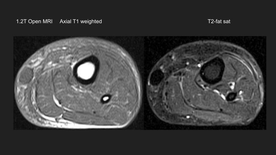
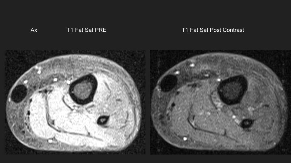
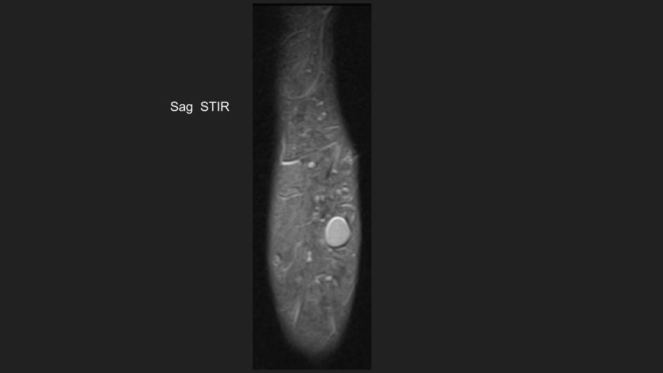
40 yr old male “injected his calved with liquid to make them bigger” 10
years ago. Now complains of bilateral calf pain.
These are images from an Open 1.2T MRI
I don’t understand the signal characteristics of the rounted foci …the
largest in the subcutaneous soft tissues of the posteromedial calf.
Intermediate T1, Dark T1 and T2 fat sat, but Bright STIR.
There is no enhancement.
How can it be dark on T2FS and Bright on STIR?
[image: 40M 10yrs post calf injections.jpg]
[image: 40M 10yrs post calf injections (1).jpg]
[image: 40M 10yrs post calf injections (2).jpg]
5 thoughts on “Calf MRI Please Help”
ocad-msk
Hi Hilary,
That’s the appearance of free silicon with some smaller nodules in the calf muscles. Here’s a couple from a butt augmentation with free silicon. PD fatsat on the left, STIR on the right.
Mark Awh
Sent: Tuesday, March 8, 2022 1:49 PM
40 yr old male “injected his calved with liquid to make them bigger” 10 years ago. Now complains of bilateral calf pain.
These are images from an Open 1.2T MRI
I don’t understand the signal characteristics of the rounted foci …the largest in the subcutaneous soft tissues of the posteromedial calf.
Intermediate T1, Dark T1 and T2 fat sat, but Bright STIR.
There is no enhancement.
How can it be dark on T2FS and Bright on STIR?
hilary.umans
I didn’t make myself clear. I understand these are Free Silicone
injections.
I don’t understand the Signal…although it is typical.
Why would it be uniformly dark on T2FS and Bright on STIR?
Thanks.
Hilary
[gallery]
hilary.umans
Joseph Zerr answered my question, thank you!
The TI peaks for fat and silicone are close, but not identical. Water based
STIR will still have signal in silicone. A separate silicone based STIR is
used in breast imaging to help find free silicone. That sequence completely
nulls the signal from silicone.
Hope that helps,
Joe
[gallery]
hilary.umans
David Wayne Robinson elaborated on the answer to my question:
To expound a little on that answer, it is necessary to remember how
spectral fat saturation differs from inversion recovery.
Specifically, spectral fat saturation takes advantage of the different
proton resonant frequencies of materials (namely water and fat), which are
measured in chemical shift parts per million (water = 4.7 ppm and fat = 1.3
ppm). The resonant frequency of injected silicon depends on the actual
compound, but as an example, the silicon compound used in ophthalmic
injections is PDMS and has a resonant frequency of 0.33 ppm. A spectral sat
band that includes fat will typically overlap with silicon.
In contrast, inversion recovery takes advantage of the different TI of
materials. And while fat and silicon-compounds have similar proton resonant
frequencies, their T1 relaxation times differ substantially. Fat is about
260 ms at 1.5T, whereas PDMS is 716 ms at 1.5T. So an IR timed for fat will
nullify fat and materials with similar and shorter T1 times. Materials
with a longer T1 times (such as silicon) will have measurable signal.
I know this is a very “physic-y” answer, but you asked a physics questions.
I hope this helps.
-David
[gallery]
avneesh.chhabra
Great explanation!
Enjoy silicon in left eye- behaves differently – see attached
btw- there are silicon maps available in spectral ct as well
David Wayne Robinson elaborated on the answer to my question:
To expound a little on that answer, it is necessary to remember how spectral fat saturation differs from inversion recovery.
Specifically, spectral fat saturation takes advantage of the different proton resonant frequencies of materials (namely water and fat), which are measured in chemical shift parts per million (water = 4.7 ppm and fat = 1.3 ppm). The resonant frequency of injected silicon depends on the actual compound, but as an example, the silicon compound used in ophthalmic injections is PDMS and has a resonant frequency of 0.33 ppm. A spectral sat band that includes fat will typically overlap with silicon.
In contrast, inversion recovery takes advantage of the different TI of materials. And while fat and silicon-compounds have similar proton resonant frequencies, their T1 relaxation times differ substantially. Fat is about 260 ms at 1.5T, whereas PDMS is 716 ms at 1.5T. So an IR timed for fat will nullify fat and materials with similar and shorter T1 times. Materials with a longer T1 times (such as silicon) will have measurable signal.
I know this is a very “physic-y” answer, but you asked a physics questions.
I hope this helps.
-David
40 yr old male “injected his calved with liquid to make them bigger” 10 years ago. Now complains of bilateral calf pain.
These are images from an Open 1.2T MRI
I don’t understand the signal characteristics of the rounted foci …the largest in the subcutaneous soft tissues of the posteromedial calf.
Intermediate T1, Dark T1 and T2 fat sat, but Bright STIR.
There is no enhancement.
How can it be dark on T2FS and Bright on STIR?
[40M 10yrs post calf injections.jpg]
[40M 10yrs post calf injections (1).jpg]
[40M 10yrs post calf injections (2).jpg]


That’s the appearance of free silicon with some smaller nodules in the calf muscles. Here’s a couple from a butt augmentation with free silicon. PD fatsat on the left, STIR on the right.
Mark Awh
Sent: Tuesday, March 8, 2022 1:49 PM
40 yr old male “injected his calved with liquid to make them bigger” 10 years ago. Now complains of bilateral calf pain.
These are images from an Open 1.2T MRI
I don’t understand the signal characteristics of the rounted foci …the largest in the subcutaneous soft tissues of the posteromedial calf.
Intermediate T1, Dark T1 and T2 fat sat, but Bright STIR.
There is no enhancement.
How can it be dark on T2FS and Bright on STIR?
injections.
I don’t understand the Signal…although it is typical.
Why would it be uniformly dark on T2FS and Bright on STIR?
Thanks.
Hilary
[gallery]
The TI peaks for fat and silicone are close, but not identical. Water based
STIR will still have signal in silicone. A separate silicone based STIR is
used in breast imaging to help find free silicone. That sequence completely
nulls the signal from silicone.
Hope that helps,
Joe
[gallery]
To expound a little on that answer, it is necessary to remember how
spectral fat saturation differs from inversion recovery.
Specifically, spectral fat saturation takes advantage of the different
proton resonant frequencies of materials (namely water and fat), which are
measured in chemical shift parts per million (water = 4.7 ppm and fat = 1.3
ppm). The resonant frequency of injected silicon depends on the actual
compound, but as an example, the silicon compound used in ophthalmic
injections is PDMS and has a resonant frequency of 0.33 ppm. A spectral sat
band that includes fat will typically overlap with silicon.
In contrast, inversion recovery takes advantage of the different TI of
materials. And while fat and silicon-compounds have similar proton resonant
frequencies, their T1 relaxation times differ substantially. Fat is about
260 ms at 1.5T, whereas PDMS is 716 ms at 1.5T. So an IR timed for fat will
nullify fat and materials with similar and shorter T1 times. Materials
with a longer T1 times (such as silicon) will have measurable signal.
I know this is a very “physic-y” answer, but you asked a physics questions.
I hope this helps.
-David
[gallery]
Enjoy silicon in left eye- behaves differently – see attached
btw- there are silicon maps available in spectral ct as well
Best!
AC
Avneesh Chhabra, M.D. M.B.A.
Professor, Radiology & Orthopedic Surgery
Chief, Musculoskeletal Radiology
Department of Radiology
5323 Harry Hines, Blvd. Dallas, Tx-75390-9316
Office: 214-648-2122
http://www.utsouthwestern.edu/radiology<http://www.utsouthwestern.edu/education/medical-school/departments/radiology/>
EXTERNAL MAIL
David Wayne Robinson elaborated on the answer to my question:
To expound a little on that answer, it is necessary to remember how spectral fat saturation differs from inversion recovery.
Specifically, spectral fat saturation takes advantage of the different proton resonant frequencies of materials (namely water and fat), which are measured in chemical shift parts per million (water = 4.7 ppm and fat = 1.3 ppm). The resonant frequency of injected silicon depends on the actual compound, but as an example, the silicon compound used in ophthalmic injections is PDMS and has a resonant frequency of 0.33 ppm. A spectral sat band that includes fat will typically overlap with silicon.
In contrast, inversion recovery takes advantage of the different TI of materials. And while fat and silicon-compounds have similar proton resonant frequencies, their T1 relaxation times differ substantially. Fat is about 260 ms at 1.5T, whereas PDMS is 716 ms at 1.5T. So an IR timed for fat will nullify fat and materials with similar and shorter T1 times. Materials with a longer T1 times (such as silicon) will have measurable signal.
I know this is a very “physic-y” answer, but you asked a physics questions.
I hope this helps.
-David
40 yr old male “injected his calved with liquid to make them bigger” 10 years ago. Now complains of bilateral calf pain.
These are images from an Open 1.2T MRI
I don’t understand the signal characteristics of the rounted foci …the largest in the subcutaneous soft tissues of the posteromedial calf.
Intermediate T1, Dark T1 and T2 fat sat, but Bright STIR.
There is no enhancement.
How can it be dark on T2FS and Bright on STIR?
[40M 10yrs post calf injections.jpg]
[40M 10yrs post calf injections (1).jpg]
[40M 10yrs post calf injections (2).jpg]
[gallery]